
In the early days of hip arthroscopy, many procedures were performed simply to find out what was happening inside the hip joint. X-Rays do not allow one to see damaged soft tissues and MR scanning was in its infancy. Even now, despite the use of specialist scans, it is still not always possible to say what is going on within a hip joint unless hip arthroscopy is undertaken.
However, in broad overview, the indications for hip arthroscopy are many and varied but perhaps may be outlined as follows:
- 1. Impingement
- 2. Articular cartilage (gristle) problems
- 3. Labral injuries
- 4. Loose body retrieval
- 5. Synovitis (inflamed hip lining)
- 6. Torn ligamentum teres (major hip ligament tear)
- 7. Hip instability
- 8. Infection
- 9. Painful joint replacement or resurfacing
- 10. Avascular necrosis (blood supply problem to ball of hip joint)
- 11. Muscle injuries around the hip (gluteus medius, gluteus minimus, gluteus maximus, rectus femoris)
- 12. Trochanteric bursitis (pain on the outside of the hip – usually in females)
- 13. Nerve release around the hip
- 14. Snapping hip
- 15. Iliopsoas tendonitis
- 16. Hip tumours
- 17. Hip fracture or hip dislocation
- 18. Foreign body retrieval (e.g. bullet)
- 19. Osteoarthritis
- 20. Diagnostic hip arthroscopy
This list is not exhaustive and it should be remembered that the results of hip arthroscopy depend in large part on the condition which is being treated. For example, we know that 80% of patients can feel better after hip arthroscopic surgery for impingement.
However, this figure can drop as low as 40% if hip arthroscopy is undertaken for osteoarthritis. Please also remember that there is no operation on the planet that can offer a 100% chance of success.
 |
 |
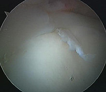
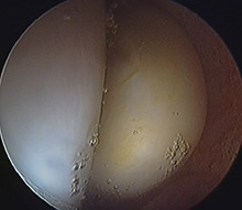
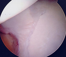
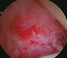
A torn acetabular labrum |
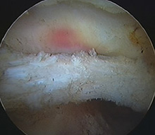
Iliopsoas tendonitis |
 |
 |
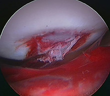
Ruptured ligamentum teres |
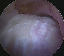
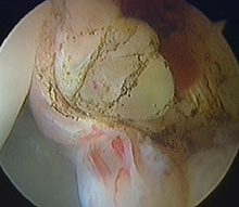
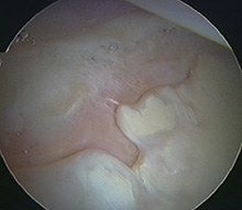
Large impingement lesion |
 |
 |
Cyst trying to burst out of the labrum (intralabral cyst) |
Lipoma (a benign tumour) of the hip joint |
 |
 |
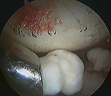
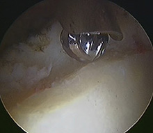
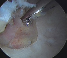
Removing a loose body from the hip |
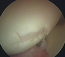
Osteoarthritis of the hip – the large lump of bone to the right was dramatically reducing hip movement until it was removed arthroscopically |
 |
 |
Fracture of the acetabulum (hip socket) |
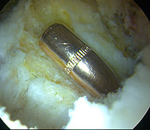
A bullet in the hip! |
 |
 |
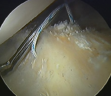
Performing a microfracture of an area of articular cartilage (gristle) loss |
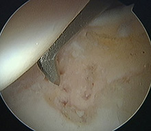
Dealing with pincer impingement – burring away part of the acetabular margin |
 |
 |
Synovitis – inflammation of the hip lining |
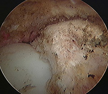
Bare bone exposed due to osteoarthritis |
 |
 |
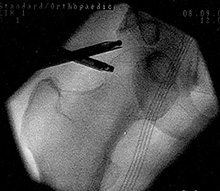
X-Ray (image intensifier) view of hip arthroscopic surgery in progress |
An area of gristle loss (osteochondral fracture) of the femoral head (hip ball) |
 |
 |
Removing osteoarthritis (central osteophyte) from the acetabulum (hip socket) |
Repairing the labrum |
The Princess Grace Hospital, 30 Devonshire Street, London, W1G 6PU | Enquiries: +44 (0)207 908 3777 | Fax: +44 (0)203 752 6501
enquiries@villarbajwa.com
© Villar Bajwa Practice (2015) | Terms & Conditions